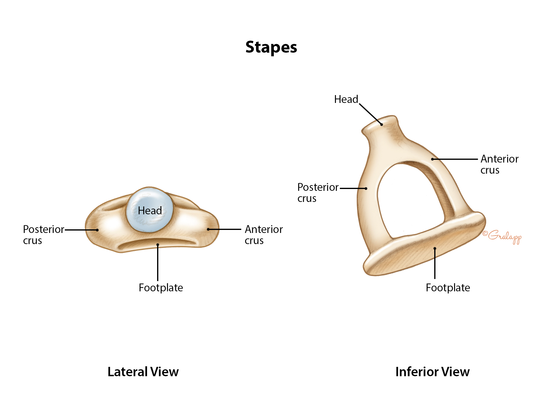
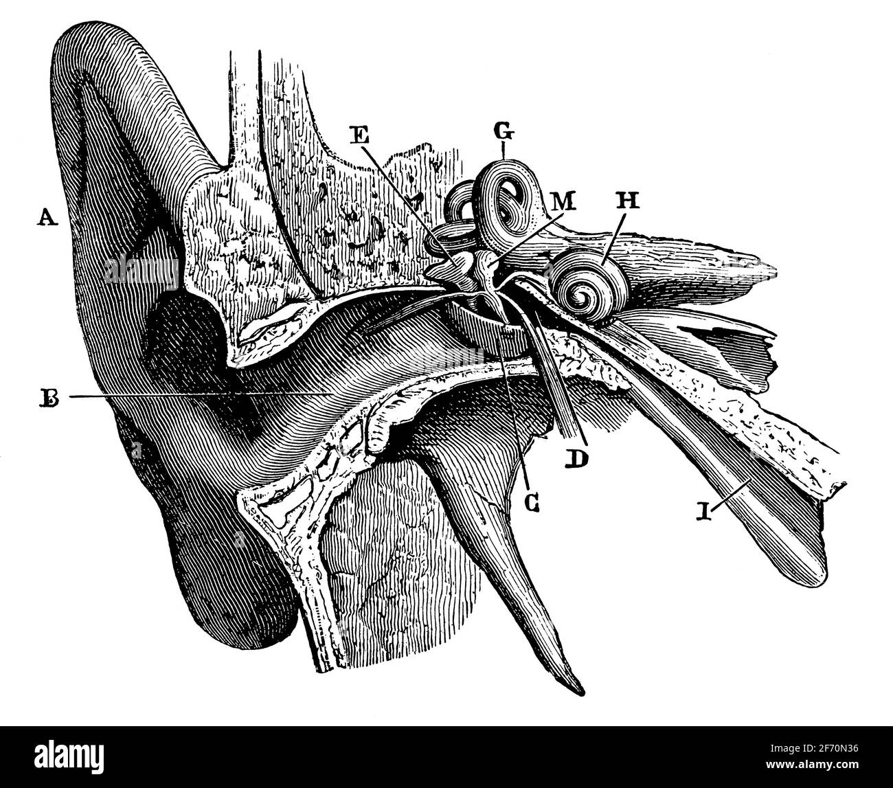
Illustration of stapes morphology following the nomenclature of
Descrição
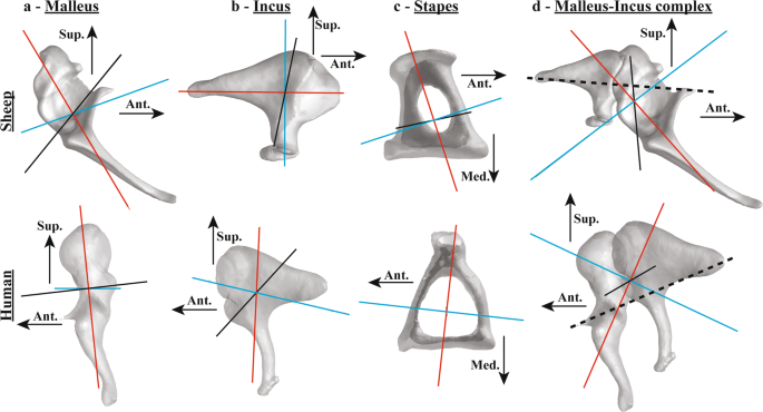
Comparison of sheep and human middle-ear ossicles: anatomy and inertial properties

Left ectotympanic (gray) and malleus (blue) in medial (A, C) and
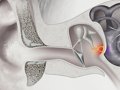
Middle Ear Contents
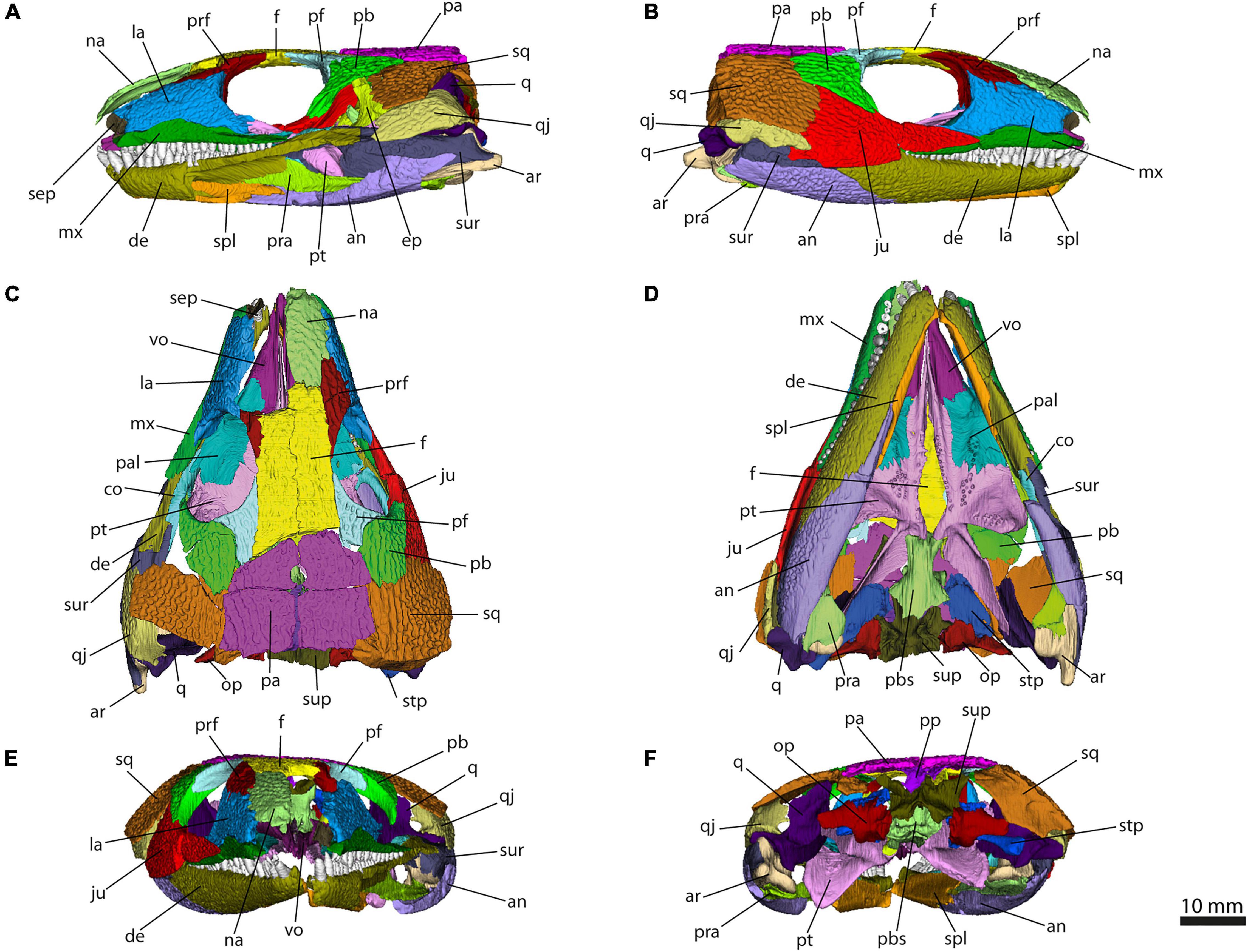
Frontiers Skull Sutures and Cranial Mechanics in the Permian Reptile Captorhinus aguti and the Evolution of the Temporal Region in Early Amniotes

Secondary Tympanic Membrane - an overview
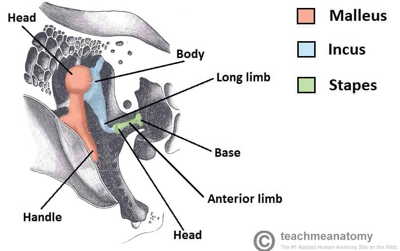
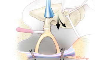
The Middle Ear - Parts - Bones - Muscles - TeachMeAnatomy
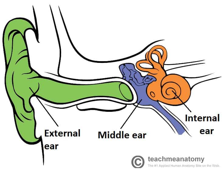
The Middle Ear - Parts - Bones - Muscles - TeachMeAnatomy
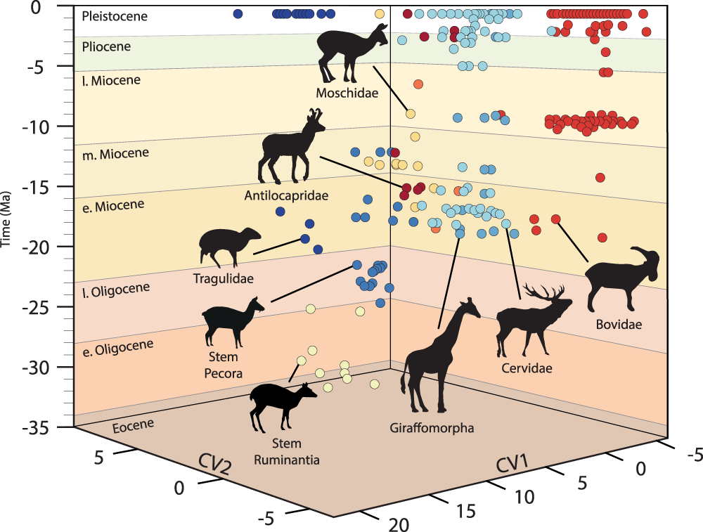
Ruminant inner ear shape records 35 million years of neutral evolution

Full article: Computed tomography and three-dimensional reconstruction of the skull of the stem tetrapod Crassigyrinus scoticus Watson, 1929
Tympanic Black and White Stock Photos & Images - Alamy

Major evolutionary transitions and innovations: the tympanic middle ear Philosophical Transactions of the Royal Society B: Biological Sciences

Mitosis - Wikipedia
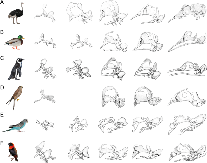
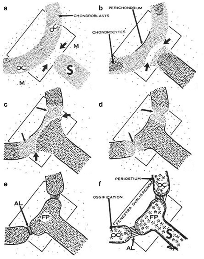
Evolution and development of the bird chondrocranium, Frontiers in Zoology
Comparative anatomy study hi-res stock photography and images - Page 12 - Alamy
de
por adulto (o preço varia de acordo com o tamanho do grupo)