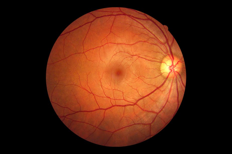
Figure 1. [The normal human retina fundus]. - Webvision - NCBI
Descrição
The normal human retina fundus photo shows the optic nerve (right), blood vessels and the position of the fovea (center).
![Figure 1. [The normal human retina fundus]. - Webvision - NCBI](https://pub.mdpi-res.com/symmetry/symmetry-15-01631/article_deploy/html/images/symmetry-15-01631-g007.png?1692867492)
Symmetry, Free Full-Text
![Figure 1. [The normal human retina fundus]. - Webvision - NCBI](https://pub.mdpi-res.com/symmetry/symmetry-15-01631/article_deploy/html/images/symmetry-15-01631-g002.png?1692867486)
Symmetry, Free Full-Text
![Figure 1. [The normal human retina fundus]. - Webvision - NCBI](https://webvision.med.utah.edu/wp-content/uploads/2019/07/KrizajFigure4sure.jpg)
What is glaucoma? by David Krizaj – Webvision
![Figure 1. [The normal human retina fundus]. - Webvision - NCBI](https://media.springernature.com/m685/springer-static/image/art%3A10.1186%2Fs12877-021-02009-z/MediaObjects/12877_2021_2009_Fig1_HTML.png)
Association of reduced retinal arteriolar tortuosity with depression in older participants from the Northern Ireland Cohort for the Longitudinal Study of Ageing, BMC Geriatrics
![Figure 1. [The normal human retina fundus]. - Webvision - NCBI](http://eyerounds.org/atlas/LARGE/Normal-fundus-LRG.jpg)
Atlas Entry - Situs Inversus of the Retinal Vessels
![Figure 1. [The normal human retina fundus]. - Webvision - NCBI](https://www.ncbi.nlm.nih.gov/corehtml/pmc/pmcgifs/bookshelf/thumbs/th-webvision-lrg.png)
Facts and Figures Concerning the Human Retina - Webvision - NCBI Bookshelf
![Figure 1. [The normal human retina fundus]. - Webvision - NCBI](https://www.ncbi.nlm.nih.gov/books/NBK11556/bin/factsf6.gif)
Facts and Figures Concerning the Human Retina - Webvision - NCBI Bookshelf
![Figure 1. [The normal human retina fundus]. - Webvision - NCBI](https://ars.els-cdn.com/content/image/1-s2.0-S266646902300026X-gr2.jpg)
Cell death mechanisms in retinal phototoxicity - ScienceDirect
![Figure 1. [The normal human retina fundus]. - Webvision - NCBI](https://www.ncbi.nlm.nih.gov/books/NBK11533/bin/sretinaf16.gif)
Simple Anatomy of the Retina - Webvision - NCBI Bookshelf
![Figure 1. [The normal human retina fundus]. - Webvision - NCBI](https://media.springernature.com/lw685/springer-static/image/chp%3A10.1007%2F978-3-030-43395-6_21/MediaObjects/107245_3_En_21_Fig1_HTML.png)
Retinal Bioengineering
![Figure 1. [The normal human retina fundus]. - Webvision - NCBI](https://www.pnas.org/cms/10.1073/pnas.2307380120/asset/a3533755-1d49-4826-ba92-7697defec4a7/assets/images/large/pnas.2307380120fig08.jpg)
Cellular migration into a subretinal honeycomb-shaped prosthesis for high-resolution prosthetic vision
![Figure 1. [The normal human retina fundus]. - Webvision - NCBI](https://media.springernature.com/m685/springer-static/image/art%3A10.1186%2Fs13024-023-00655-y/MediaObjects/13024_2023_655_Fig4_HTML.png)
Retinal ganglion cell repopulation for vision restoration in optic neuropathy: a roadmap from the RReSTORe Consortium, Molecular Neurodegeneration
de
por adulto (o preço varia de acordo com o tamanho do grupo)