Figure 3 from Stimulated Emission Depletion (STED) Microscopy: from Theory to Practice
Descrição
Figure 3. Fluorescence depletion of two common dyes in STED microscopy, Atto647N (black, diamonds) and Atto655 (red, circles), as a function of the depletion laser intensity. Error bars for Atto647N appear smaller than the point size of the average value. - "Stimulated Emission Depletion (STED) Microscopy: from Theory to Practice"
![Figure 3 from Stimulated Emission Depletion (STED) Microscopy: from Theory to Practice](https://zeiss-campus.magnet.fsu.edu/tutorials/superresolution/stedconcept/stedconcepttutorialfigure1.jpg)
ZEISS Microscopy Online Campus, Interactive Tutorials
![Figure 3 from Stimulated Emission Depletion (STED) Microscopy: from Theory to Practice](https://www.frontiersin.org/files/Articles/641330/fchem-09-641330-HTML/image_m/fchem-09-641330-g002.jpg)
Frontiers Shedding New Lights Into STED Microscopy: Emerging
![Figure 3 from Stimulated Emission Depletion (STED) Microscopy: from Theory to Practice](https://ars.els-cdn.com/content/image/1-s2.0-S0030401817305138-gr2b.jpg)
Aberrations in stimulated emission depletion (STED) microscopy
![Figure 3 from Stimulated Emission Depletion (STED) Microscopy: from Theory to Practice](https://onlinelibrary.wiley.com/cms/asset/224189e0-ff8d-45b6-9e66-e895933f6983/jnc13257-fig-0003-m.jpg)
Stochastic optical reconstruction microscopy (STORM) in comparison
![Figure 3 from Stimulated Emission Depletion (STED) Microscopy: from Theory to Practice](https://pub.mdpi-res.com/biosensors/biosensors-11-00330/article_deploy/html/images/biosensors-11-00330-g001-550.jpg?1631517552)
Biosensors, Free Full-Text
![Figure 3 from Stimulated Emission Depletion (STED) Microscopy: from Theory to Practice](https://www.mdpi.com/biosensors/biosensors-11-00330/article_deploy/html/images/biosensors-11-00330-g002-550.jpg)
Biosensors, Free Full-Text
![Figure 3 from Stimulated Emission Depletion (STED) Microscopy: from Theory to Practice](https://royalsocietypublishing.org/cms/asset/759f606c-8349-4019-bcd4-cb063dadb42c/rstb20130597f03.jpg)
Dissecting tripartite synapses with STED microscopy
![Figure 3 from Stimulated Emission Depletion (STED) Microscopy: from Theory to Practice](https://royalsocietypublishing.org/cms/asset/d9b97b06-0d11-4614-910b-b81fb9eaaad3/rstb20130597f02.jpg)
Dissecting tripartite synapses with STED microscopy
![Figure 3 from Stimulated Emission Depletion (STED) Microscopy: from Theory to Practice](https://www.researchgate.net/publication/268290268/figure/fig2/AS:295355901071367@1447429742620/Determination-of-the-STED-microscopes-resolution-a-STED-left-half-and-confocal.png)
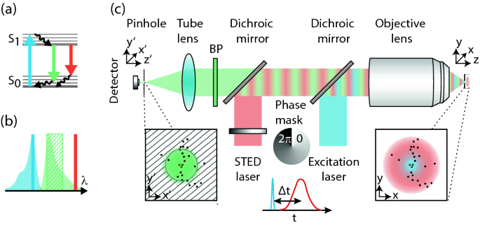
Determination of the STED microscope's resolution. (a) STED (left
![Figure 3 from Stimulated Emission Depletion (STED) Microscopy: from Theory to Practice](https://www.picoquant.com/?ACT=32&f=sted-setup.jpg&fid=71&d=7356)
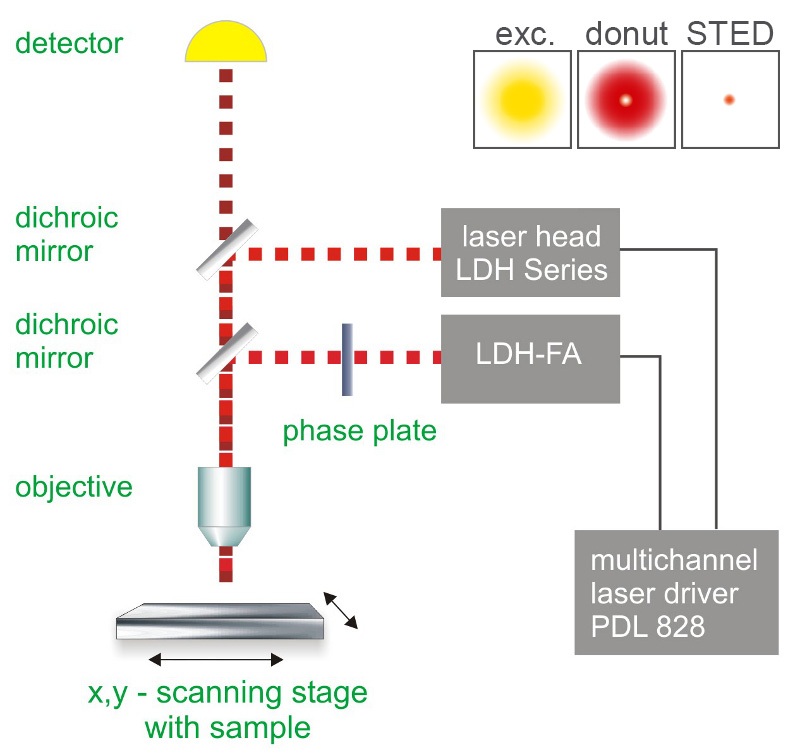
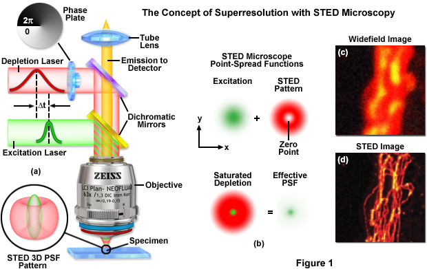
Stimulated Emission Depletion Microscopy (STED)
![Figure 3 from Stimulated Emission Depletion (STED) Microscopy: from Theory to Practice](https://media.springernature.com/lw685/springer-static/image/chp%3A10.1007%2F978-3-030-34413-9_1/MediaObjects/455781_1_En_1_Fig9_HTML.png)
STED Nanoscopy
![Figure 3 from Stimulated Emission Depletion (STED) Microscopy: from Theory to Practice](https://d3i71xaburhd42.cloudfront.net/4bf45c891c77216fc88e21bdd41979908abbcf70/6-Table1-1.png)
Figure 3 from Stimulated Emission Depletion (STED) Microscopy
![Figure 3 from Stimulated Emission Depletion (STED) Microscopy: from Theory to Practice](https://abberior.rocks/wp-content/uploads/STED-PAINT_DNA-PAINT_principle.jpg)
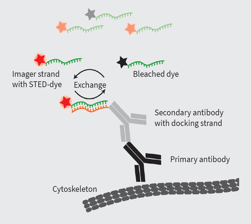
STED-PAINT for high-performance superresolution
de
por adulto (o preço varia de acordo com o tamanho do grupo)